 English
 Français
Medical optical imaging
Medical optical imaging includes the acquisition and display of human body images, at various scales, produced by a number of physical phenomena such as photon absorption, emission, scattering and reflection. All biological tissues exhibit optical properties. For example, all molecules absorb several wavelengths ; choosing the proper wavelengths allows selecting the molecules to be imaged. Images are obtained by processing the data supplied by the tissue optical properties. These imaging techniques aimed at acquiring anatomical, functional, molecular and metabolic information in order to contribute to the understanding of living tissues, healthy or pathological, in an interventional or diagnostic purpose.
We are interested in the wavelength range including UV, visible, near-, mid- , far-infrared and TeraHerz. In this spectral range, optical imaging has numerous advantages. Indeed, in addition to the selectivity mentioned above, optical imaging does not use ionizing radiation or radioactive elements ; the technique employs non invasive processes which can be repeated safely and allows to differentiate biological structures on the basis of their chemical composition. Moreover, the technologies used lead to instruments exhibiting smaller sizes and lower costs than CT (computed tomography), MRI, PET … Some of these devices are well adapted for a use at the point of care.
Over the last years, medical optical imaging is constantly evolving and new technologies have arisen resulting in medical devices already on the market and many others at an advanced development stage.
Since the ophtamologists’ slit-lamp, otorhinolaryngologists’ laryngoscope, urologists’ first rigid endoscopes, a huge amount of progress has been achieved, particularly with the advent of optical fibers, lasers, miniaturized cameras, display screens and image processing algorithms.
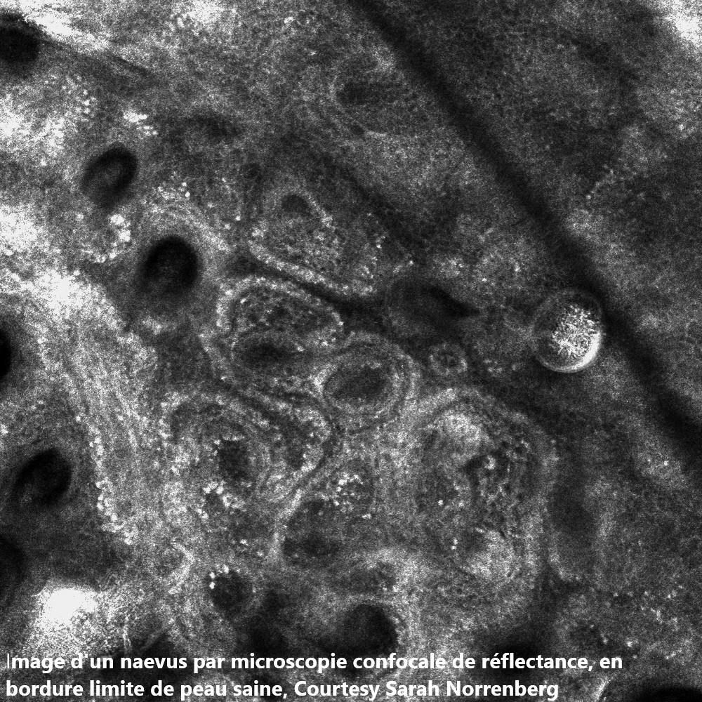
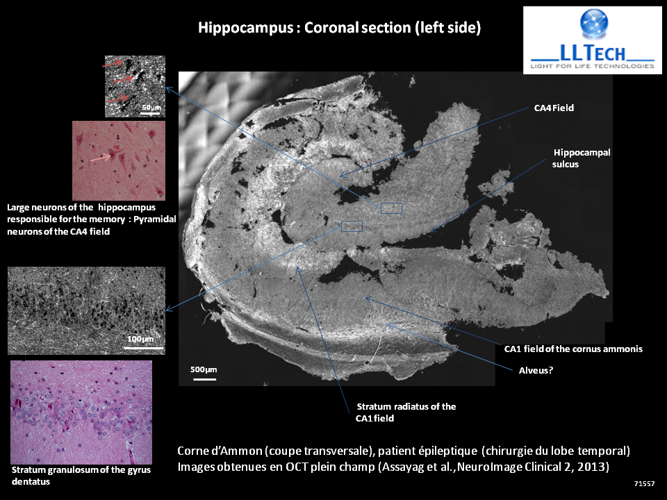
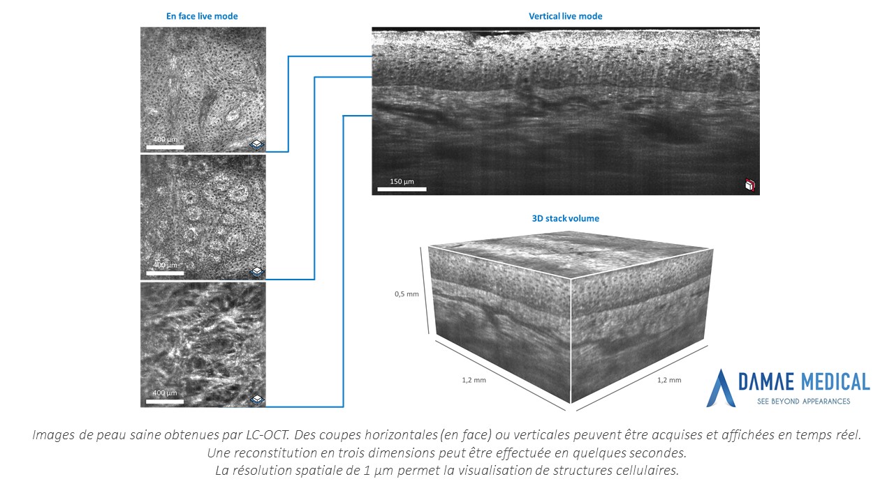
The most outstanding recent achievements include among others: fluorescence imaging, OCT (optical coherent tomography), confocal endomicroscopy, photo-acoustic imaging, polarimetric imaging, Raman spectroscopy, DRS (diffuse reflectance spectroscopy), multiphotonic imaging, DOT (diffuse optical tomography), hyperspectral imaging …
Optical biopsy has become a reality in a number of medical fields.
Optical imaging is now a major component of image guided surgery and theranostic development. Augmented reality and deep learning are today’s technological advances which lead and continue to lead to improved images and perceptions.

- See French version
- See French version
- Line-field confocal optical coherence tomography for high-resolution noninvasive imaging of skin tumors
Arnaud Dubois, Olivier Levecq, Hicham Azimani, David Siret, Anaïs Barut, Mariano Suppa, Véronique del Marmol, Josep Malvehy, Elisa Cinotti, Pietro Rubegni, Jean-Luc Perrot
Accéder au fichier PDF
- Photoacoustic tomography: principles and advances
Jun Xia, Junjie Yao and Lihong V. Wang
Accéder au fichier PDF
- Photoacoustic microscopy and computed tomography: from bench to bedside
Lihong V. Wang and Liang Gao
Accéder au fichier PDF
- A Practical Guide to Photoacoustic Tomography in the Life Sciences
Lihong V. Wang and Junjie Yao
Accéder au fichier PDF
- Photoacoustic Tomography: In Vivo Imaging from Organelles to Organs
Lihong V. Wang and Song Hu
Accéder au fichier PDF
Upcoming content